Journal of Endocrinology and Metabolism, ISSN 1923-2861 print, 1923-287X online, Open Access
Article copyright, the authors; Journal compilation copyright, J Endocrinol Metab and Elmer Press Inc
Journal website http://www.jofem.org

Original Article

Volume 7, Number 1, February 2017, pages 5-17


Modified Fine-Needle Aspiration Biopsy for Calcitonin, Procalcitonin and Carcinoembryonic Antigen Levels in the Diagnosis of Thyroid Nodules With Medullary Thyroid Carcinoma

Figures

Figure 1.
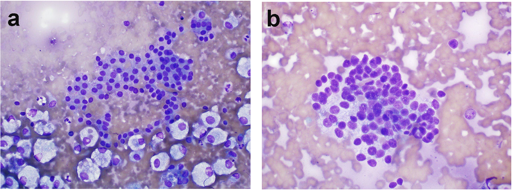
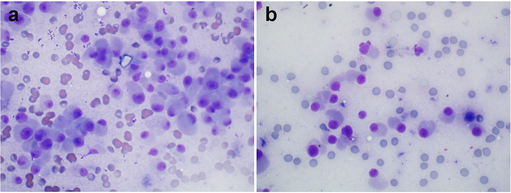
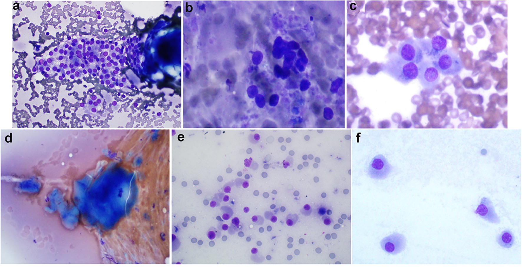
Figure 1. (Patient 1) (a) Results of FNB biopsy of the right thyroid nodules (two nodules, 1a and 1b biopsied together): follicular cells trying to form microfollicles. Follicular cells show some degree of atypia. These features suggest follicular lesions of undetermined significance (Diff-Quik, × 600). (b) Results of FNB biopsy of the left thyroid nodule (nodule 2a): follicular cells forming microfollicles. Follicular cells show some degree of atypia. These features suggest follicular lesions of undetermined significance (Diff-Quik, × 600). (c) Results of MFNB biopsy of the right thyroid nodules (two nodules, 1a and 1b biopsied together): cellular smears consisting of discohesive cells with abundant amphophillic cytoplasm, and eccentrically placed nuclei with prominent nucleoli. These features along with elevated serum calcitonin suggest medullary thyroid cancer (Diff-Quik, × 600). (d) MFNB of the left thyroid nodule (nodule 2a): amorphous material suggestive of amyloid material (Congo red stain not done) (Diff-Quik, × 600). (e) MFNB of the left thyroid nodule (nodule 2a): crowded groups of mildly enlarged cells with oval to focally spindled nuclei, moderate to abundant cytoplasm and finely stippled chromatin. While not classic for medullary carcinoma, the concurrent finding of elevated serum calcitonin makes the diagnosis more likely (Diff-Quik, × 600). (f) MFNB of the left thyroid nodule (nodule 2a): cells with oval to focally spindled nuclei, abundant cytoplasm and finely stippled chromatin. While not classic for medullary carcinoma, the concurrent finding of elevated serum calcitonin makes the diagnosis more likely (Diff-Quik, × 600). FNB: fine-needle biopsy; MFNB: modified fine-needle biopsy; Diff. quick: Diff quick stain.
Figure 2.
Figure 2. (Patient 2) (a) Results of FNB biopsy of the right thyroid nodule: follicular cells forming microfollicles and showing some degree of atypia. These features suggest follicular lesions of undetermined significance (Diff-Quik, × 600). (b) Results of MFNB of right thyroid nodule: crowded groups of mildly enlarged cells with oval to focally spindled nuclei, moderate cytoplasm and finely stippled chromatin. While not classic for medullary carcinoma, the concurrent finding of elevated serum calcitonin makes this diagnosis more likely (Diff-Quik, × 600). FNB: fine-needle biopsy; MFNB: modified fine-needle biopsy; Diff. quick: Diff quick stain.
Figure 3.
Figure 3. (Patient 3) (a) MFNB of the right thyroid nodule showing: cellular smears consisting of discohesive cells with abundant amphophillic cytoplasm, and eccentrically placed nuclei with prominent nucleoli (oncocytic features) (Diff-Quik, × 600). (b) MFNB of the left thyroid nodule: smears consisting of discohesive, focally enlarged cells with eccentric nuclei with occasional nucleoli, coarse chromatin and abundant cytoplasm. These features along with elevated serum calcitonin suggest medullary thyroid cancer (Diff-Quik, × 600). FNB: fine-needle biopsy; MFNB: modified fine-needle biopsy; Diff. quick: Diff quick stain.

Tables

Table 1. Serum Levels of Calcium, Calcitonin (Ct), Procalcitonin (PCt) and Carcinoembryonic Antigen (CEA) in Patients With Medullary Thyroid Cancer Following Intravenous Administration of Calcium Gluconate
 
Serum levelsMinutes post-calcium administration
0251015
Calcium gluconate (2.5 mg/kg) was administered intravenously over 1 min and blood samples were obtained at the indicated various time intervals as mentioned in “Methods” [22]. Reference values: basal serum Ct 0.0 - 8.4 pg/mL, basal PCt 0.00 - 0.50 ng/mL, CEA 0.2 - 4.7 ng/mL. The serum Ct threshold for the identification of MTC was defined a > 26 pg/mL for basal levels in females [22]. The threshold for calcium-stimulated Ct level was > 79 pg/mL in females [22]. The threshold for calcium-stimulated PCt level was > 1.65 ng/mL. The threshold for stimulated CEA was considered as 17.16 ng/mL.
Calcium (mg/dL)
  Patient 19.510.611.110.79.6
  Patient 29.911.611.210.89.8
  Patient 39.010.410.910.99.4
Calcitonin (pg/mL)
  Patient 141555642889718
  Patient 2587814212382
  Patient 36943932018498
Procalcitonin (ng/mL)
  Patient 10.322.323.991.781.05
  Patient 22.486.306.977.114.88
  Patient 30.421.453.941.011.38
Carcinoembryonic antigen (ng/mL)
  Patient 12.502.812.322.002.86
  Patient 21.861.761.751.691.83
  Patient 33.224.103.843.463.09


Table 2. Calcitonin (Ct), Procalcitonin (PCt) and Carcinoembryonic Antigen (CEA) Levels in Serum and Fine-Needle Aspiration Biopsy (FNB) Samples in Patients With Medullary Thyroid Cancer
 
Tumor markersSerumFNB aspirate
Levels of Ct, PCt and CEA, in fine-needle aspirates (FNB) were determined as mentioned in “Methods”. Blood was drawn just prior to performing FNB for the determination of serum levels of Ct, PCt and CEA. Patient 1 had three nodules and all three nodules were biopsied. Rt thyroid nodule, 1a and the adjacent second Rt nodule, 1b were biopsied together and considered as a single aspirate sample as mentioned in “Methods”. In this patient, Lt thyroid nodule, 2a was also biopsied. In patient 2, the right lobe nodule,1a was biopsied. Patient 3 had bilateral thyroid nodules which were biopsied (Rt thyroid nodule,1a Lt thyroid nodule, 2b) as outlined in “Methods”. Reference ranges: basal serum Ct 0.0 - 8.4 pg/mL, basal PCt 0.00 - 0.50 ng/mL, CEA 0.2 - 4.7 ng/mL. The basal serum Ct threshold for the identification of MTC was defined as > 26 pg/mL for females [22]. The threshold for calcium-stimulated Ct level of > 1,000 pg/mL in needle biopsy samples was considered diagnostic for MTC [24]. The diagnostic values for FNB aspirate samples for PCt and CEA were > 2.0 and > 18 ng/mL, respectively.
Calcitonin (pg/mL)
  Patient 148726 (Rt thyroid nodule-1a and 1b)
824 (Lt thyroid nodule-2a)
  Patient 2521,996 (Rt thyroid nodule-1a)
  Patient 370744 (Rt thyroid nodule-1a)
1,120 (Lt thyroid nodule-2a)
Procalcitonin (ng/mL)
  Patient 10.921.6 (Rt thyroid nodule-1a and 1b)
2.0 (Lt thyroid nodule-2a)
  Patient 23.24.4 (Rt thyroid nodule-1a)
  Patient 30.611.8 (Rt thyroid nodule-1a)
8.0 (Lt thyroid nodule-2b)
Carcinoembryonic antigen (ng/mL)
  Patient 12.913.9 (Rt thyroid nodule-1a and 1b)
14.2 (Lt thyroid nodule-2a)
  Patient 216621.6 (Rt thyroid nodule-1a)
  Patient 3417.7 (Rt thyroid nodule-1a)
19.6 (Lt thyroid nodule-2b)


Table 3. Calcitonin (Ct), Procalcitonin (PCt) and Carcinoembryonic Antigen (CEA) Levels in Serum and Modified Fine-Needle Aspiration Biopsy (MFNB) Samples in Patients With Medullary Thyroid Cancer
 
Tumor markersSerumMFNB aspirate
Levels of Ct, PCt and CEA, in modified fine-needle aspirates (MFNB) were determined as mentioned in “Methods”. Blood was drawn just prior to performing MFNB for the determination of serum levels of Ct, PCt and CEA. Patient 1 had three nodules and all three nodules were biopsied. Rt thyroid nodule, 1a and the adjacent second Rt nodule, 1b were biopsied together and considered as a single aspirate sample as mentioned in “Methods”. In this patient, Lt thyroid nodule, 2a was also biopsied. In patient 2, the right lobe nodule, 1a was biopsied. Patient 3 had bilateral thyroid nodules which were biopsied (Rt thyroid nodule, 1a and Lt thyroid nodule, 2b) as outlined in “Methods”. Reference ranges: basal serum Ct 0.0 - 8.4 pg/mL, basal PCt 0.00 - 0.50 ng/mL, CEA 0.2 - 4.7 ng/mL. The basal serum Ct threshold for the identification of MTC was defined as > 26 pg/mL for females [22]. The threshold for Ct level of > 1,000 pg/mL in needle biopsy samples was considered diagnostic for MTC [24]. The diagnostic values for MFNB aspirate samples for PCt and CEA were > 2.0 and > 18 ng/mL.
Calcitonin (pg/mL)
  Patient 1521,036 (Rt thyroid nodules-1a and 1b)
1,870 (Lt thyroid nodule-2a)
  Patient 2681,440 (Rt thyroid nodule-1a)
  Patient 3821,341 (Rt thyroid nodule-1a)
1,328 (Lt thyroid nodule-2a)
Procalcitonin (ng/mL)
  Patient 10.483.2 (Rt thyroid nodule-1a and 1b)
2.8 (Lt thyroid nodule-2a)
  Patient 22.407.8 (Rt thyroid nodule 1a)
  Patient 30.436.8 (Rt thyroid nodule-1a)
9.2 (Lt thyroid nodule-2b)
Carcinoembryonic antigen (ng/mL)
  Patient 13.121.9 (Rt thyroid nodule (1a and 1b)
18.6 (Lt thyroid nodule-2a)
  Patient 217029.9 (Rt thyroid nodule-1a)
  Patient 33.652.3 (Rt thyroid nodule-1a)
28.1 (Lt thyroid nodule-2b)


Table 4. Serum Levels of Calcium, Calcitonin (Ct), Procalcitonin (PCt) and Carcinoembryonic Antigen (CEA) in Patients With Benign Thyroid Nodules (n = 3, Patients 1, 2 and 4) and Multifocal Papillary Thyroid Cancer (n = 1, Patient 3) Following Intravenous Administration of Calcium Gluconate
 
Serum levelsMinutes post-calcium stimulation
0251015
Calcium gluconate (2.5 mg/kg) was administered intravenously over 1 min and blood samples were obtained at the indicated various time intervals as mentioned in “Methods”. Reference ranges: basal serum Ct 0.0 - 8.4 pg/mL, basal PCt 0.00 - 0.50 ng/mL, CEA 0.2 - 4.7 ng/mL. The serum Ct threshold for the identification of MTC was defined as > 26 pg/mL for basal levels in females [22]. The threshold for calcium-stimulated Ct levels was > 79 pg/mL in females [22]. In this study, the basal serum threshold for PCt was defined as > 0.5 ng/mL and the threshold for calcium-stimulated abnormal PCt level was defined as > 1.65 ng/mL. The threshold for basal CEA was > 5.0 ng/mL and the threshold for stimulated CEA was considered as 17.16 ng/mL.
Calcium (mg/dL)
  Patient 18.610.110.09.19.3
  Patient 29.210.410.39.49.6
  Patient 38.610.09.69.69.5
  Patient 48.910.610.19.89.4
Calcitonin (pg/mL)
  Patient 130125884730
  Patient 239112975844
  Patient 32.93624174.2
  Patient 42756484222
Procalcitonin (pg/mL)
  Patient 10.140.150.130.120.13
  Patient 20.050.050.0050.050.05
  Patient 30.160.130.150.170.18
  Patient 40.180.100.050.050.05
Carcinoembryonic antigen (ng/mL)
  Patient 10.240.230.200.250.21
  Patient 21.61.51.61.51.5
  Patient 31.71.61.21.41.5
  Patient 40.80.91.00.450.67


Table 5. Calcitonin (Ct), Procalcitonin (PCt), and Carcinoembryonic Antigen (CEA) in Serum and Modified Find-Needle Aspiration Biopsy (MFNB) Samples in Benign Thyroid Nodules (n = 3, Patients 1 - 3) and in Another Patient (Patient 4) With Subcentimeter Multicentric Papillary Thyroid Cancer
 
SerumMFNB aspirate
Blood was drawn for the determination of Ct, PCt, and CEA levels just prior to performing MFNB. In patients 1 - 3 only MFNB was performed and the aspirate results are shown above. In patient 4, initially FNB was performed which showed abnormal cells suggestive of follicular neoplasm and in this patient the FNB aspirate samples showed low values of Ct, PCt, and CEA (data not shown). In this patient MFNB was performed subsequently and the cytology report was consistent with papillary thyroid cancer and the MFNB results are shown in this Table. Reference ranges: basal serum Ct 0.0 - 8.4 pg/mL, basal PCt 0.00 - 0.50 ng/mL, CEA 0.2 - 4.7 ng/mL. The MTC diagnostic value for basal serum Ct level was > 26 pg/mL for females [22]. In this study, the basal serum threshold for PCt was defined as > 0.5 ng/mL and for CEA this value was > 5.0 ng/mL. The threshold for Ct level of > 1,000 pg/mL in needle biopsy samples was considered diagnostic for MTC [24]. The diagnostic values for MFNB aspirate samples for PCt and CEA were > 2.0 and > 18 ng/mL, respectively.
Calcitonin (pg/mL)
  Patient 1283.2
  Patient 244< 0.05
  Patient 34.71.9
  Patient 4332.7
Procalcitonin (ng/mL)
  Patient 10.120.22
  Patient 20.410.43
  Patient 30.360.19
  Patient 40.180.38
Carcinoembryonic antigen (ng/mL)
  Patient 12.93.7
  Patient 22.22.4
  Patient 31.33.2
  Patient 44.13.6