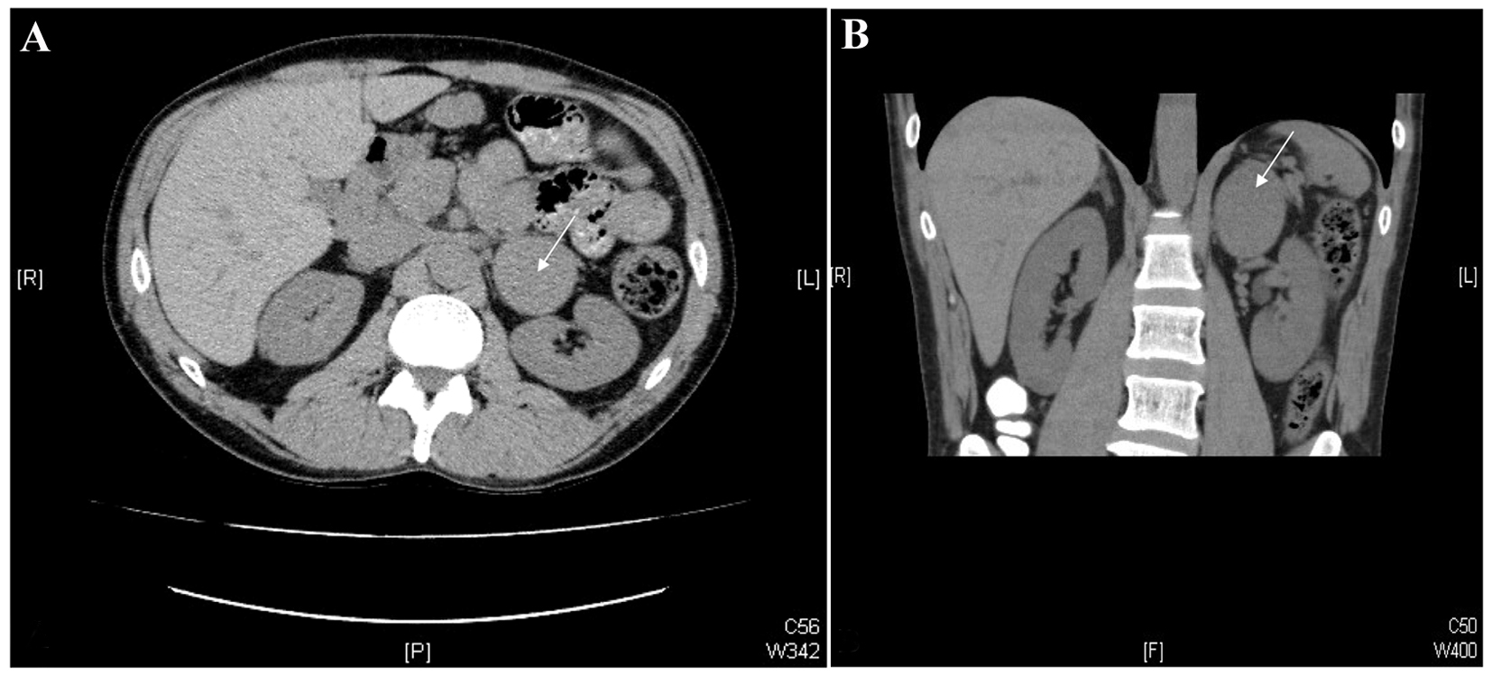
Figure 1. CT images of the abdomen. Axial (A) and coronal (B) images from a non-contrast-enhanced CT scan of the abdomen. Note the 5.8× 5.0 cm mass in the left adrenal gland (white arrows).
| Journal of Endocrinology and Metabolism, ISSN 1923-2861 print, 1923-287X online, Open Access |
| Article copyright, the authors; Journal compilation copyright, J Endocrinol Metab and Elmer Press Inc |
| Journal website http://www.jofem.org |
Case Report
Volume 4, Number 1-2, April 2014, pages 39-46
An Unusual Case of a Composite Pheochromocytoma With Neuroblastoma
Figures
Tables
| Test | Result | Reference range |
|---|---|---|
| Epinephrine | 742 nmol | < 100 nmol/day |
| Norepinephrine | 589 nmol | < 500 nmol/day |
| Dopamine | 3,179 nmol | < 2,600 nmol/day |
| Normetanephrine | 7.3 µmol | < 3.3 µmol/day |
| Metanephrine | 19.9 µmol | < 1.7 µmol/day |
| VMA | 70 µmol | 6 - 36 µmol/day |
| Test | Result | Reference range |
|---|---|---|
| Chromogranin A | 242 U/L | < 21 U/L |
| Plasma free metanephrine | 3.34 nmol/L | < 0.50 nmol/L |
| Plasma free normetanephrine | 6.09 nmol/L | < 0.90 nmol/L |