Journal of Endocrinology and Metabolism, ISSN 1923-2861 print, 1923-287X online, Open Access
Article copyright, the authors; Journal compilation copyright, J Endocrinol Metab and Elmer Press Inc
Journal website http://www.jofem.org

Case Report

Volume 2, Number 4-5, October 2012, pages 187-189


Acute Pancreatitis as the First Manifestation of Parathyroid Adenoma

Figures

Figure 1.
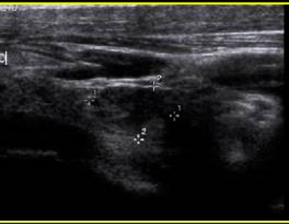
Figure 1. Head and Neck Ultrasound showed 1.5 × 1× 1.1 cm oval solid mass at the posterior inferior aspect of the right thyroid lobe.
Figure 2.
Figure 2. Following administration of 26.3 mCi 99m-technetium labeled Cardiolite, planar and SPECT images of the parathyroid gland were obtained. Immediate images (a) demonstrate a focal area of activity in the right lobe projected over the right lobe of the thyroid which persists on delayed image (b). Findings are consistent with parathyroid adenoma.
Figure 3.
Figure 3. Hypercellular parathyroid gland consistent with adenoma. Nests of endocrine cells without significant atypia, occasional small cystic spaces and gland-like areas.