


| Journal of Endocrinology and Metabolism, ISSN 1923-2861 print, 1923-287X online, Open Access |
| Article copyright, the authors; Journal compilation copyright, J Endocrinol Metab and Elmer Press Inc |
| Journal website http://www.jofem.org |
Letter to the Editor
Volume 8, Number 1, February 2018, pages 13-14
Usefulness of Scintigraphy to Evaluate Adrenal Tumors
Tadanao Harigaea, b, Naonori Tsudaa, b, Hiroki Hashimotoa, Hidekatsu Yanaia, c
aDepartment of Internal Medicine, National Center for Global Health and Medicine Kohnodai Hospital, Chiba, Japan
bBoth authors equally contributed to this work.
cCorresponding Author: Hidekatsu Yanai, Department of Internal Medicine, National Center for Global Health and Medicine Kohnodai Hospital, 1-7-1 Kohnodai, Ichikawa, Chiba 272-8516, Japan
Manuscript submitted January 19, 2018, accepted January 30, 2018
Short title: Scintigraphy to Evaluate Adrenal Tumors
doi: https://doi.org/10.14740/jem488w
| To the Editor | ▴Top |
If we find adrenal tumors in patients, how should we do? We should examine to make clear whether the tumor is hormone-producing or not, and whether the tumor is benign or malignant. To make sure that the tumor is malignant or not, what can we do? The diameter of the tumor can be useful. Tumor size at presentation (mean diameter at diagnosis > 10 cm) has been reported to be the most important indicator of malignancy [1]. Another review article described that a functioning adrenal carcinoma should be considered when an adrenal mass measures more than 5 cm in diameter [2]. However, in the previous study which examined adrenal tumors of 57 patients who underwent adrenalectomy, the mean diameter of the resected primary adenocarcinomas was 3.0 and 4.5 cm, respectively [3]. A very recent study reported that tumor size ≥ 40 mm was significantly associated with malignancy [4]. In addition to tumor size, elevated serum dehydroepiandrosterone sulphate (DHEAS) can predict malignancy [4].
After the rule-out of the possibility for malignancy using CT, MRI and measurement of serum DHEAS, we examine blood hormone levels. Plasma renin and aldosterone levels, serum ACTH, cortisol, and catecholamine levels (dopamine, noradrenaline and adrenaline) are measured. If cortisol increased and ACTH decreased, Cushing’s syndrome was suspected. In Cushing’s syndrome, diurnal variation of cortisol disappeared and urinary cortisol level increased (> 80 µg/day). I-adosterol scintigraphy can confirm the existence of cortisol-producing tumor (Fig. 1). A patient B was referred to our hospital due to resistant hypertension. Serum noradrenaline level was high (576 pg/mL; normal range, 100 - 450 pg/mL); however, daily urinary metanephrine and normetanephrine levels were within normal range. Therefore, we performed I-metaiodobenzylguanidine (MIBG) scintigraphy, and could detect the uptake of I-MIBG in adrenal tumor (Fig. 1), which made us diagnose this patient.
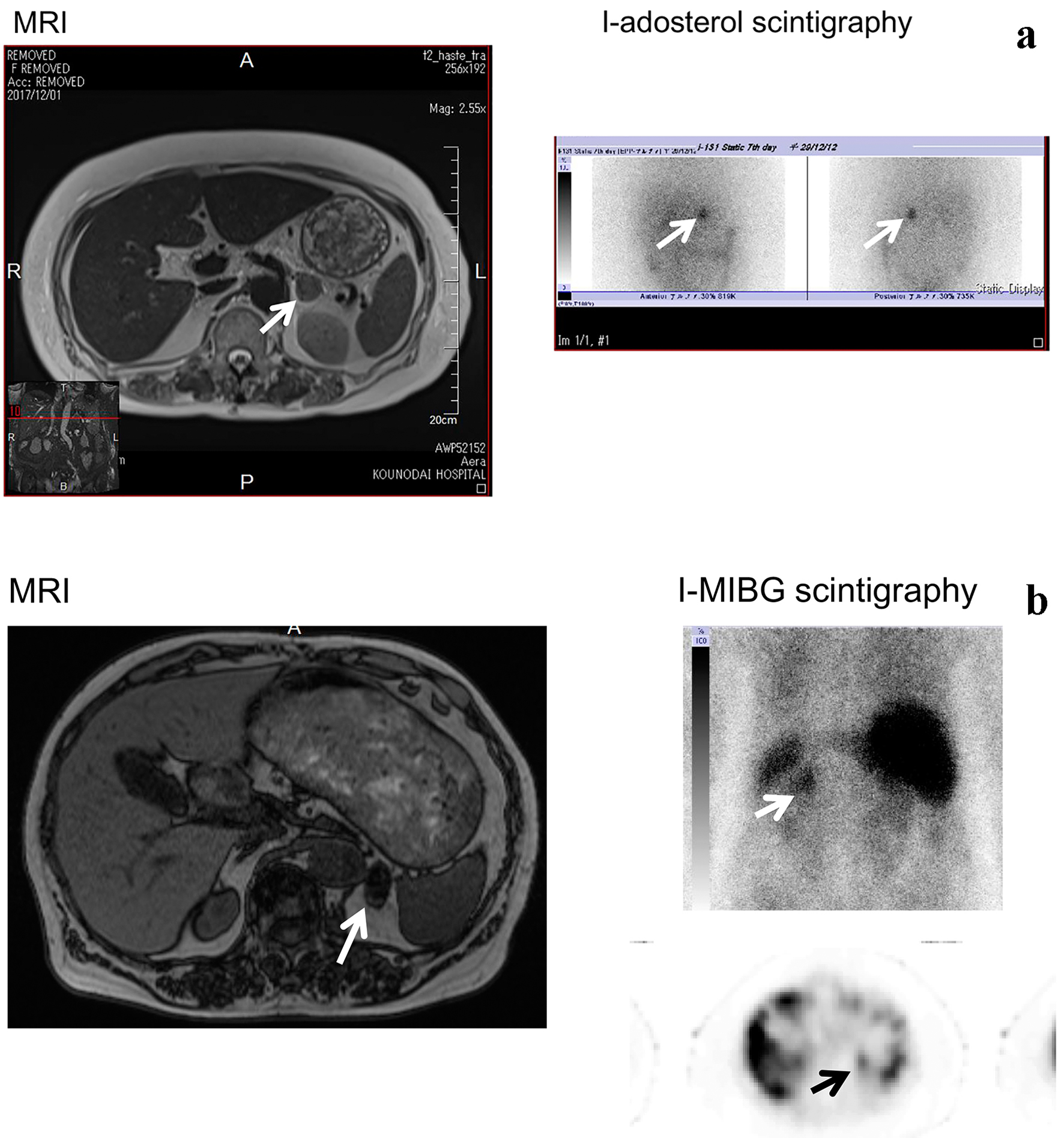 Click for large image | Figure 1. MRI and scintigraphy of patients with Cushing’ syndrome (a) and pheochromocytoma (b). |
To evaluate functioning (hormone-producing) adrenal tumors, in addition to endocrinological tests, an appropriate scintigraphy may be useful.
Conflict of Interest
The authors declare that they have no conflict of interest concerning this article.
| References | ▴Top |
- Allolio B, Hahner S, Weismann D, Fassnacht M. Management of adrenocortical carcinoma. Clin Endocrinol (Oxf). 2004;60(3):273-287.
doi - Iihara M, Obara T. [Diagnosis and surgical treatment of adrenal tumors]. Nihon Geka Gakkai Zasshi. 2005;106(8):479-483.
pubmed - Linos DA, Stylopoulos N, Raptis SA. Adrenaloma: a call for more aggressive management. World J Surg. 1996;20(7):788-792; discussion 792-783.
- Foo E, Turner R, Wang KC, Aniss A, Gill AJ, Sidhu S, Clifton-Bligh R, et al. Predicting malignancy in adrenal incidentaloma and evaluation of a novel risk stratification algorithm. ANZ J Surg. 2017.
doi
This article is distributed under the terms of the Creative Commons Attribution Non-Commercial 4.0 International License, which permits unrestricted non-commercial use, distribution, and reproduction in any medium, provided the original work is properly cited.
Journal of Endocrinology and Metabolism is published by Elmer Press Inc.